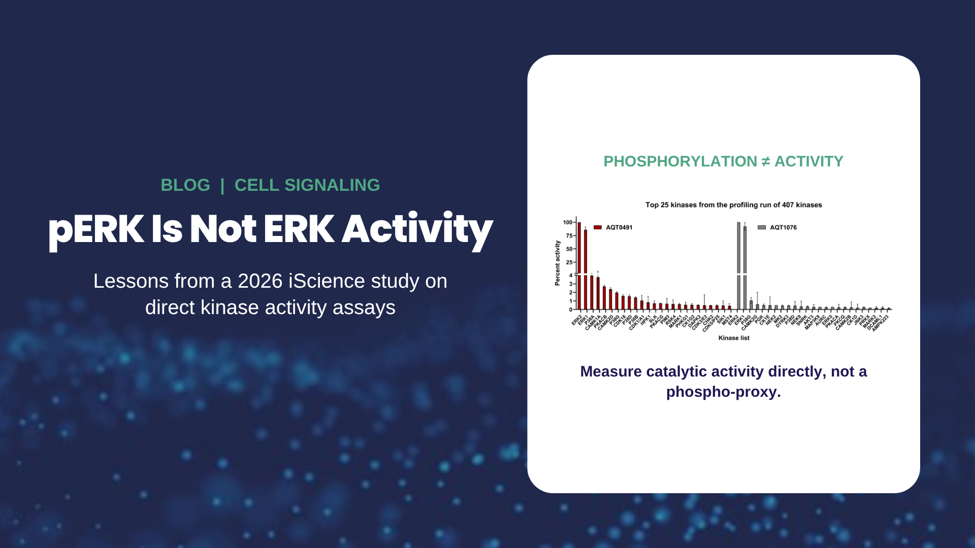
If you treat cancer cells with an ERK1/2 inhibitor and probe a western blot for phospho-ERK, you might expect the signal to drop. In many cases it goes up instead. Does that mean the drug failed? An independent 2026 study from David Engelberg's lab at the Hebrew University of Jerusalem, published in iScience, answers that question directly, and the answer has implications for anyone who uses phosphorylation as a stand-in for kinase activity.
Key takeaways
- Five ERK1/2 inhibitors (BVD523/ulixertinib, GDC0994/ravoxertinib, SCH772984, ASN007, and temuterkib) all caused strong, sustained phosphorylation of ERK at its activating TEY motif.
- Direct measurement of ERK catalytic activity in cell lysates showed activity was suppressed, even while phospho-ERK rose.
- The rise in phospho-ERK reflects the inhibitors cutting off ERK's own negative feedback on the upstream pathway, not a failure of inhibition.
- The authors conclude that a direct, specific kinase activity assay, not pTEY abundance, should be used to judge ERK's catalytic status.
The paradox: inhibited ERK that looks "more active"
ERK1 and ERK2 sit at the bottom of the RTK–Ras–Raf–MEK cascade, which is constitutively active in many cancers. That makes ERK an attractive drug target. But ERK does two jobs at once: it drives proliferation, and it also restrains the upstream pathway through negative feedback. Block ERK, and that brake comes off.
The researchers tested five clinical-stage ERK1/2 inhibitors in cells transformed by intrinsically active ERK1 variants and in nine cancer-derived cell lines. Every inhibitor produced dramatic, sustained phosphorylation of ERK (and of MEK), consistent with release of feedback inhibition. Read on its own, that western blot would suggest the pathway was more active than before treatment.
What direct activity measurement showed
To find out what ERK was actually doing, the team measured ERK catalytic activity directly in lysates using the PhosphoSens® ERK1/2 Cell Lysate Kinase Assay, a continuous, fluorescence-based assay that tracks phosphorylation of a selective sensor peptide in real time.
- Transformed cells: after just 2 hours of BVD523 treatment, ERK activity was already significantly reduced in all of the transformed clones, even as TEY phosphorylation climbed.
- Cancer cell lines: in MDA-MB-231 cells, which have high basal ERK phosphorylation, ERK activity was high and was efficiently inhibited by both BVD523 and SCH772984, with SCH772984 the more efficient inhibitor. In HeLa cells, ERK activity was barely detectable with or without inhibitor.
In other words, phosphorylation and catalytic activity told opposite stories. The authors put it plainly: a direct and specific kinase assay "should be the sole readout for the status of Erks' catalytic capabilities."
What else the study found
The activity data were one part of a broader picture. All five inhibitors arrested cells in G1 and activated the stress kinase p38, a response more typical of cytotoxic therapy than targeted drugs. Some cell viability persisted even after 72 hours, and phosphoproteomic analysis of BVD523-treated cells revealed large-scale changes, most prominently in Rho signaling. Because inhibited ERK remains heavily phosphorylated, the authors suggest it may be primed for rapid reactivation once the drug is gone, and they argue that ERK inhibitors should be combined with agents that prevent pathway reactivation.
Why this matters beyond ERK
Phospho-specific antibodies are the default readout for kinase pathway activity in many labs. They are useful, but they report a modification state at a single moment, not catalytic turnover. Whenever feedback loops, inhibitor binding modes, or scaffold interactions decouple phosphorylation from activity, a phospho-blot can point you in the wrong direction. That risk shows up in several common situations:
- Mechanism-of-action studies: confirming a compound actually shuts down catalytic output, rather than inferring it from a phospho-signal that feedback can push either way.
- Pharmacodynamic and target-engagement studies: tracking pathway suppression, rebound, or incomplete engagement after dosing.
- Resistance and model comparisons: comparing functional activity across cell lines, mutants, or patient-derived models under identical conditions, without re-optimizing an antibody for each.
Measuring kinase activity directly in lysates
PhosphoSens-Lysate assays measure endogenous kinase activity in crude cell or tissue lysates with no antibodies and no wash steps. A kinase-selective sensor peptide fluoresces as it is phosphorylated, so each well produces a real-time progress curve on a standard fluorescence plate reader. In addition to ERK1/2, validated lysate assays are available for AKT1/2/3, GSK3α/β, DNA-PK, JNK1/2/3, p38α/β/γ/δ, CDK1/2/3/5, CDK2/Cyclin E1, and ZAP70.
If you are relying on phospho-ERK to judge inhibitor activity, see the ERK1/2 lysate assay or talk to an AssayQuant scientist →
Reference
Darash-Yahana M, Soudah N, Baskin A, Levy-Saar I, Levitzki A, Engelberg D. Erk inhibitors intercept Erk-mediated negative feedback while imposing cell-cycle arrest and activating p38. iScience. 2026;29(7):116629. doi:10.1016/j.isci.2026.116629
This study was conducted independently by the Engelberg laboratory using a commercially available AssayQuant kit.